

Description
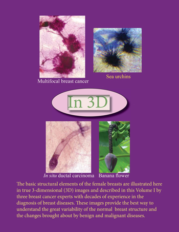
These volumes have inbuilt stereoscopic glasses with the help of which you can see real three-dimensional histopathology images of the facing page.
Dr. László Tabár and his colleagues have published a new, groundbreaking book in 3D! It is titled “Understanding the Breast in Health and Disease”. It is available in both English and Spanish.
The purpose of this book is two-fold:
To educate physicians, members of the diagnostic (radiologists specialized in breast imaging and pathologists specialized in breast pathology) and therapeutic teams(surgeons, medical and radiation oncologists), that combining the large thin section (10X8 cm) histology technique with the subgross, 3-D technique facilitates better understanding of normal and pathologic breast tissue, and makes precise correlation with the radiologic imaging methods a reality. Regular mammography screening of asymptomatic women – the prerequisite for a significant decrease in morality from breast cancer – has added a new dimension to the traditional interaction between pathologists and radiologists. The imager can recognize the structural elements, which are vividly visualized on the subgross (3-D) histology images. This technique serves to bridge the gap that separates the small but high resolution, 4 micron histopathology image from the comprehensive, but low resolution radiology imaging techniques. It is the large, thick section (3D) histology technique that brings the radiologist and pathologist to a common ground for a better understanding of breast morphology.
 To educate women about the normal anatomy of the breast, its many variations, demonstrate and provide explanation for most of the non-malignant breast changes that may cause clinical symptoms such as breast pain, serous or bloody nipple discharge, how breast cysts develop, what is fibrocystic change / fibroadenoma / radial scar and other benign breast changes. We do not attempt to explain cellular details, instead, we will deal with the subgross anatomy of the most important structural elements:
- The lobules, where the milk is produced and where most of the breast cancers originate. Its size varies between 0.7-1.5 mm and it has the appearance of “gloves” with many fingers. The number of “fingers” (so-called acini or ductules) vary depending on age, phase in the menstrual cycle, pregnancy, lactation, etc. The lobules and the very last segment of the milk duct (the terminal duct) build a functional unit, called the terminal ductal lobular unit, TDLU.
- The milk ducts and their branches.
- The adipose tissue
- Fibrosis.
 Our aim is to explain how the TDLUs and the milk ducts become distended and distorted by the accumulation of fluid or cancer cells. We also intend to explain the revolutionary changes brought about by the development of modern breast imaging methods, how early detection of breast cancer saves lives, one of the great accomplishments of cancer research during the past forty years.
The book also comes with detailed commentaries from Dr. Tabár that you can listen to while going through the book.