Content & Methodology
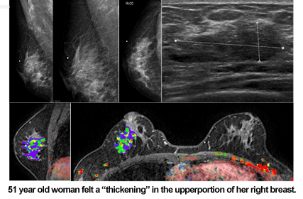
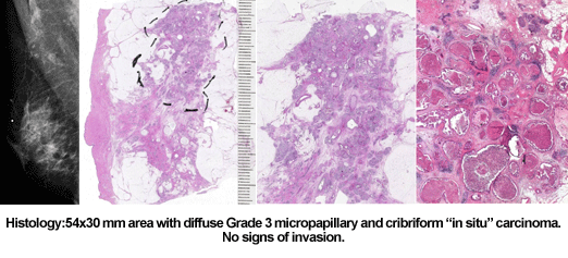
Illustration of the entire spectrum of breast pathology – correlated to mammography/US/MRI – using:
- Large histology sections (10 x 8 cm).
- Thick section / 3D pathology.
- The corresponding mammography/US/MRI images.
- Didactic correlation between the clinical picture, mammograms, galactogram, and thick section / 3D histology is an approach which can be readily understood and applied in practice.


- The mammographic, ultrasound, MRI, and histologic images of a wide range of breast diseases will be explained using the latest presentation technology tools.
- In his past lecture series, Dr. Tabár used thousands of 35mm slides and drew illustrations on yellow transparencies that were projected on overhead. These effective albeit cumbersome tools were his format for training more than fifteen thousand medical professionals over a span of sixteen years.
- The recent developments of revolutionary computer technology, such as Microsoft’s TabletPC initiative and “digital ink”-capable PowerPoint software, have enabled Dr. Tabár to considerably improve his ability to communicate with the audience. High-resolution scanned images of his highly appreciated teaching slides are now displayed in innovative PowerPoint presentations. Also, the use of TabletPC technology makes it possible both to draw directly onto the PowerPoint slides and produce stand-alone hand-drawn illustrations.
- These new tools have enabled Dr. Tabár to create a completely new series of presentations and replace the outdated analog lecture tools.
